LMI
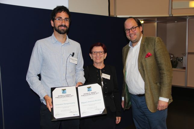
Miguel and Chandini Win Bernd R. Binder Publication Prize 2017. Congrats!
Read more: https://www.technoclone.com/en/brb-award

Recent News
6 years, 2 months ago
Comments Off on Miguel and Chandini Win Bernd R. Binder Publication Prize 2017
LMI wins the Alexander Schmidt Award at the Annual Meeting of the Society of Thrombosis and Hemostasis Research (GTH) in Vienna.
>> http://www.gth2018.org/
>> http://viennamotion.zenfolio.com/p102480911/ea4924a5c
Miguel and Chandini publish Report in Science! Congrats!
Title: “Host DNases prevent vascular occlusion by neutrophil extracellular traps”
Weblink: http://science.sciencemag.org/content/358/6367/1202
Positions for PhD and Postdoctoral Fellows are available on a rolling basis to highly motivated candidates with the following qualifications:
• Master or diploma in biochemistry, cell biology, immunology, or a related field
• Strong academic record and knowledge in biochemistry, cell biology, and immunology
• Strong interest in inflammation, innate and adaptive immunity, and vascular biology
• Experience in basic science research and translational medicine
• Experience in murine disease models and FELASA certification is a plus
• Proficiency in English is required
• PhD in inflammation, immunity, or related field (Postdoc applicants only)
• 1+ first author publication in a top-tier journal (Postdoc applicants only)
Please submit your curriculum vitae, a statement of your research interests, and contact information of two academic references in electronic form to:
Tobias Fuchs, PhD
Principal Investigator
Laboratory of Molecular Inflammation
University Medical Center Hamburg-Eppendorf
Institute of Clinical Chemistry and Laboratory Medicine
Martinistraße 52, 20246 Hamburg, Germany
E-Mail: fuchs@inflammation.de
Web: www.inflammation.de
Dr. Boettcher and LMI publish article in Scientific Reports!
Title: “Therapeutic targeting of extracellular DNA improves the outcome of intestinal ischemic reperfusion injury in neonatal rats”
Weblink: https://www.nature.com/articles/s41598-017-15807-6
Abstract: Thrombosis and inflammation cooperate in the development of intestinal infarction. Recent studies suggest that extracellular DNA released by damaged cells or neutrophils in form of extracellular traps (NETs) contributes to organ damage in experimental models of ischemia-reperfusion injury. Here we compared the therapeutic effects of targeting fibrin or extracellular DNA in intestinal infarction after midgut volvulus in rats. Following iatrogenic midgut volvulus induction for 3 hours, we treated animals with a combination of tissue plasminogen activator (tPA) and low molecular weight heparin (LMWH) to target fibrin or with DNase1 to degrade extracellular DNA. The therapeutic effects of tPA/LMWH and DNase1 were analyzed after 7 days. We observed that both therapeutic interventions ameliorated tissue injury, apoptosis, and oxidative stress in the intestine. DNase1, but not tPA/LMWH, reduced intestinal neutrophil infiltration and histone-myeloperoxidase-complexes, a surrogate marker of NETs, in circulation. Importantly, tPA/LMWH, but not DNase1, interfered with hemostasis as evidenced by a prolonged tail bleeding time. In conclusion, our data suggest that the therapeutic targeting of fibrin and extracellular DNA improves the outcome of midgut volvulus in rats. DNase1 therapy reduces the inflammatory response including NETs without increasing the risk of bleeding. Thus, targeting of extracellular DNA may provide a safe therapy for patients with intestinal infarction in future.
Rachita publishes article in Frontiers of Immunology. Congrats!
Title: “Neutrophil Extracellular Traps Contain Selected Antigens of Anti-Neutrophil Cytoplasmic Antibodies.”
Weblink: https://www.frontiersin.org/articles/10.3389/fimmu.2017.00439/full
Abstract: Neutrophil extracellular traps (NETs) are chromatin filaments decorated with enzymes from neutrophil cytoplasmic granules. Anti-neutrophil cytoplasmic antibodies (ANCAs) bind to enzymes from neutrophil cytoplasmic granules and are biomarkers for the diagnosis of systemic vasculitides. ANCA diagnostics are based on indirect immunofluorescence (IIF) of ethanol-fixed neutrophils. IIF shows a cytoplasmic staining pattern (C-ANCA) due to autoantibodies against proteinase 3 (PR3) or a perinuclear staining pattern (P-ANCA) due to autoantibodies against myeloperoxidase (MPO). The distinct ANCA-staining patterns are an artifact of ethanol fixation. Here, we tested NETs as a substrate for the detection of ANCAs in human sera. We observed that P-ANCAs specifically stained NETs, while C-ANCAs targeted the cell bodies of netting neutrophils. The distinct ANCA-staining patterns were caused by the presence of MPO, but not PR3, in NETs. Using NETs as a substrate for IIF, we characterized ANCAs in sera of patients with ANCA-associated vasculitis (AAV). Furthermore, we inhibited serine proteases by diisopropylfluorophosphate to prevent chromatin unfolding and the release of NETs and thus generated neutrophils with MPO-positive nuclei and PR3-positive cytoplasm, which resembled the appearance of ethanol-fixed neutrophils. In conclusion, our data suggest that NETs are selectively loaded with antigens recognized by P-ANCAs, and netting neutrophils provide a physiological substrate for ANCA detection in patients with AAV.
Tobias Fuchs gives a talk entitled “Contribution of NETs to Thrombosis” at ISTH 2017 in Berlin.
Date: July 11th / 14:45 – 16:15
Link: http://www.isth2017.org/
Miguel publishes a review about “Circulating extracellular DNA – cause or consequence of thrombosis?” in Seminars in Thrombosis and Hemostasis (doi: 10.1055/s-0036-1597284).
© 2015 LMI – Laboratory of Molecular Inflammation | Legal Notice

